

Preserving Microvascular Health: Detecting Asymptomatic Diabetic Retinal Damage Early in Peoria and Kewanee, IL
Diabetic retinopathy often progresses silently without initial pain or noticeable visual loss. Through high-resolution digital retinal mapping and optical coherence tomography, Illinois Vision Clinic detects subtle microvascular changes early, allowing timely clinical intervention to preserve clear, functional vision for adults managing diabetes throughout Central Illinois.
Navigating health management near Grandview Drive in Peoria requires steady vigilance, particularly for individuals living with type one or type two diabetes. Elevated blood sugar levels gradually weaken the tiny blood vessels that feed the delicate retina at the back of the eye. Early structural changes occur quietly, long before fuzzy vision or dark spots appear in your visual field. Detecting microvascular lesions, which are tiny localized swellings or leaks in retinal capillaries, requires advanced diagnostic imaging beyond simple eye chart tests. Our clinical team utilizes wide-field digital imaging to capture panoramic views of the entire retinal landscape, identifying subtle vascular abnormalities in the earliest stages, when treatment is most effective.
High-Resolution Retinal Mapping via Advanced Diagnostic Scanning
Protecting permanent vision requires detailed visualization beneath the retina’s visible surface. Incorporating Optical Coherence Tomography (OCT), a high-resolution, non-invasive imaging modality that uses light waves, allows optometrists to view cross-sectional layers of retinal tissue in real time. This micro-level view reveals microscopic macular edema, fluid accumulation, or subtle cellular thickening that traditional physical examinations might miss. Early identification of capillary non-perfusion prevents irreversible tissue damage and structural swelling. Collaborative medical reporting directly to your primary care physician ensures seamless teamwork between optometric specialists and endocrinologists, aligning systemic diabetes control with proactive ocular protection.
Retinal Diagnostic Comparison: Standard Visual Check vs. Advanced Retinal Imaging
| Screening Parameter | Standard Visual Acuity Test | Wide-Field Retinal Mapping & OCT |
| Primary Focus Area | Measures central clarity on wall chart | Maps microvascular structures and tissue layers |
| Asymptomatic Disease Detection | Cannot identify early capillary leakage | Detects microscopic lesions before vision loss |
| Tissue Layer Visualization | Limited to front surface inspection | Provides cross-sectional cellular analysis |
| Long-Term Disease Tracking | Records broad changes in lens strength | Compares micro-anatomical scans annually |
Crucial Benefits of Annual Medical Retinal Screening
- Early detection of microaneurysms before measurable vision impairment occurs.
- Non-invasive cross-sectional tissue scans without physical discomfort or downtime.
- Coordinated progress reporting sent directly to primary health care practitioners.
- Personalized lifestyle and optical strategies designed to protect delicate microvasculature.
Common Questions About Diabetic Eye Exams
Managing diabetes involves monitoring numerous health markers, making annual eye exams an essential defense against permanent vision loss. Many patients are surprised to learn that significant retinal damage can develop without any physical pain or immediate blurring. Clear clinical information empowers patients to stay ahead of diabetic complications through proactive screening.
How frequently should patients diagnosed with diabetes undergo medical retinal exams?
Medical guidelines advise all diabetic individuals to complete a comprehensive dilated retinal evaluation at least once per year. Annual monitoring allows our clinicians along N. Knoxville Ave in Peoria to track microscopic vessel changes over time. Sooner follow-up visits may be scheduled if active microvascular changes require close observation.
Does a medical diabetic retinal examination differ from a routine eye test for glasses?
Routine vision tests focus exclusively on evaluating refractive errors to prescribe corrective lenses. Medical diabetic exams thoroughly evaluate the health of the internal retinal vasculature, fluid levels, and nerve layer integrity using advanced OCT technology. Comprehensive medical scanning protects long-term sight rather than simply updating lens prescriptions.
What happens if early diabetic retinal changes are detected during my visit?
Identifying subtle vascular leakage during early stages allows our optometrists to coordinate care with your managing physician immediately. Adjusting systemic blood sugar parameters combined with localized clinical monitoring often prevents further disease progression. Timely clinical management preserves structural eye health and stabilizes long-term visual performance.
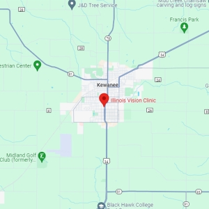
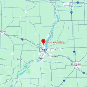
Schedule Your Diabetic Retinal Exam in Peoria or Kewanee
Protect your vision against silent diabetic microvascular changes with advanced diagnostic imaging. Contact Dr. Kerry H. Head, OD, and Dr. Ivetta Daccache, OD, FAAO at Illinois Vision Clinic today to book your comprehensive diabetic eye examination.